 by Mike Brooks |
Last Updated: December 27, 2021
by Mike Brooks |
Last Updated: December 27, 2021 3D bioprinting immersed in the 21st century. It’s a mind-boggling idea to print human tissue through additive manufacturing.
As an Amazon Associate, I earn from qualifying purchases. If you make a purchase after clicking on a link I may earn a small commission at no extra cost to you.
One can say it’s an outstanding step in tissue regenerative and tissue engineering medicine.
Over the past few decades, this technology has been used to try to create functional tissue constructs that mimic human tissue.
Quick Navigation
- What Is 3D Bioprinting?
- Why Bio Printed Tissue May Replace Animals and Humans in Preclinical Trials
- How Does 3D Bioprinting Work?
- Why 3D Bioprinting Instead of Normal Donation?
- What Is the Purpose of Three Dimensional Bioprinting?
- When Was 3D Bioprinting Invented?
- The Procedure for 3D Bioprinting
- Techniques of Bioprinting
- What Are the Disadvantages of 3D Bioprinting?
- How Many Types of Bioprinting Are There?
- Who Could Benefit From 3D Bioprinting?
- What Is 3D Bioprinting of Tissues and Organs?
- Which Material Can Be Used in Bioprinting?
- Wrap Up
Therefore, 3D bioprinting could terminate the long processes involved in animal and human clinical trials of drugs.
Plus, it could be the solution for organ shortages during organ transplants which is prone to fail because of tissue rejection. This breakthrough would end the desperate state of donating organs worldwide. Here’s all you need to know about 3D bioprinting.
What Is 3D Bioprinting?
Additive manufacturing has extended its application in organ engineering. The process involves building an organ or a tissue in layers (one layer or another). It utilizes the bottoms-up approach of 3D printing.
The layer-by-layer approach ensures you deposit primary cells, bionic and other materials in a specific fashion that mimics the typical cellular architecture.
Thus, the process leads to a synthesized tissue or an organ with the normal functionality and structure of the complex natural tissue.
In 3 Dimensional bioprinting, you print biomolecules and cells onto substrates to form a particular pattern that holds the construct together as the required 3D form. Please note that 3D bioprinting uses living human stem cells, tissues, and more.
Thus you must follow the modalities involved with living tissues. These modalities include biocompatibility of cells and materials, cell sensitivity to your printing materials and methods, perfusion, and growth factor delivery.
Why Bio Printed Tissue May Replace Animals and Humans in Preclinical Trials
The process of bioprinting is automated. Thus, this automation ensures precise cell patterns and controlled extracellular communication and organization.
Also, the layer-to-layer fabrication of the biosynthesized tissue ensures that the printed tissue has interconnected pores.
These pores are ideal for nutrients and gas perfusion. It also allows intra- and inter-cellular communication.
Therefore, the bio-printed tissue or organ that has improved intercellular and intracellular communication will be ideal for in vivo human physiology.
This feature would make the synthetic tissue better because it helps in the data obtained in the pre-clinical trials also because animal tissue may not sufficiently predict human pathophysiological response.
How Does 3D Bioprinting Work?
In the human body, tissues get damaged, and they degenerate daily. Yet, your tissue regeneration capabilities may not be sufficient in dealing with frequent trauma like accidents or heart diseases.
Over time, treating such conditions depends on tissue or organ transplantation. Thus, the entire process risks an immune response or graft rejection.
In solving the two issues, 3D printing comes in handy.
Why? Because you need an organ, the goal of regenerative medicine through 3D bioprinting is to provide your stem cells with the organ or tissue that you need. Then you’ll have the perfect tissue that does not attract these autoimmune responses.

The concept of 3D bioprinting entails material science principles and human biology for synthesizing tissues and organs.
Thus, the major focus is restoring damaged organs or tissues, like liver cirrhosis or heart failure. Hence the idea revolves around emulating the native biological complexity of the tissue leading to stem cell differentiation leading to tissue regeneration.
Why 3D Bioprinting Instead of Normal Donation?
In the usual donation, the process that leads to tissue or organ rejection results from the cell formations and connection interphase, among other things. It’s influenced by growth factors, such as vascular endothelial growth factors.
This process is somewhat random and doesn’t allow customized distribution of extracellular matrices or cells. Plus, it’s less efficient and time-consuming. From the economical and logistical viewpoint, this downside results in graft non-feasibility for the clinical application.
Thus, additive manufacturing helps explore tissue engineering through the top-down approach in 3D bioprinting.
This approach has a controlled nature in depositing matter which helps produce precise geometrics which is anatomically accurate using a computer-aided design.
What Is the Purpose of Three Dimensional Bioprinting?
According to Allevi 3D printers, over 120,000 US citizens need an organ donation.
Other innumerable patients have chronic and other terminal health conditions due to post-transplant immuno-suppression and others because of the long-term damage of the same.
Thus, the increased pressure and the need for organ transplant alternatives. Additive manufacturing has helped the scientific and the medical community to compose multidisciplinary researchers, engineers, and physicians to face challenges related to human health.
Three-dimensional bioprinting is a tool promising the elimination of organ and tissue transplant waiting lists. Also, in pharmaceutical development, bioprinting has a faster and less costly way of conducting drug clinical trials that are biologically relevant to animals and humans.
What’s more? Biomedical devices like sugar stents are a recent development because of 3D printing.
This device, for instance, helps surgeons to join veins reducing complications. Three-dimensional printing also helps to offer easier drug delivery systems.
Amazingly, further 3D bioprinting evolution will make bone tissue engineering and skin tissue, cardiac tissue, organ patches, or full organ replacement using a patient’s stem cells.
The goal of 3D printing is to provide physicians and researchers with a better tool for target treatments with improved outcomes.
When Was 3D Bioprinting Invented?
This question takes us back to the early 1900s when the dot matrix printers were discovered.
The three-dimensional printers able to print tangible objects from data by Charles Hall formed a basis for all hobbyists and engineers to print different objects, including tissues and buildings.
However, three-dimensional bioprinting started in 2000 when someone created prosthetics and implants that nearly matched the patient’s characteristics. The medical field has embraced non-biological uses of 3D printing plus anatomical modeling.
In 2003 Thomas Boland made the first three-dimensional bioprinter to print a living tissue using bio-ink of biocompatible substances. Following the 2003 breakthrough was the successful implantation of the 2006 1st lab-made human bladder and the 2009 bioprinting of the 1st blood vessel.
The Procedure for 3D Bioprinting
Three-dimensional bioprinting strategies revolve around the precise layering of materials. The bioprinting process involves preparation, printing, and post-handling phases.
In the preparation phase, you design 3D models using computer graphics. These models must be anatomically accurate.
You also select the bio-ink you will use. This selection means you determine the muscle tissue or the structure you want, thus selecting the right materials, including mammalian cells, endothelial cells, or any other cell types you need.
The second step involves selecting additive materials, and the last step involves maturing the fabricated structures.
Techniques of Bioprinting
You can carry out bioprinting either scaffold-free or scaffold-based. Scaffold-based mode; the matrix comprises the stratum used in the manufacturing process. This biomaterial matrix patterns the bio-ink. Therefore, you can use cell-laden hydrogel, a film, or a nanofiber.
Please note: the outcome biological construct must closely mimic the typical extracellular matrix environment. This aspect allows the biological constructs’ cells to increase and grow.
Scaffold-free bioprinting entails depositing tissue and cell aggregates as spheroids, cylinders, honeycombs, etc. the second process involves putting tissue spheroids into pipettes then depositing the pipette into a confined space of the 3D bioprinter mold by extrusion.
Then the cells form their cell matrix, which leads to tissue maturation; hence you eliminate the mold.
What Are the Disadvantages of 3D Bioprinting?
A contamination risk in continuous ink-jet bioprinting: the bio-ink that is not deflected in the substrate recirculates into your printer. The recirculation may lead to contamination.
Lack of bioprinting components like inadequate software can define biological molecules, biomaterials, and cell placement. This lack hampers 3D bioprinting operations.
In Scaffold deformation, your newly formed tissues can fail if you don’t provide mechanical and structural support. Thus you must manufacture stable 3d constructs.
How Many Types of Bioprinting Are There?
There are several three-dimensional printing techniques for the fabrication and selective patterning of the extracellular matrix. They include:
- Ink-jet printing
- Extrusion printing
- Laser-assisted bioprinting
- Stereolithographic bioprinting
Inkjet Based Three Dimensional Bioprinting
Ink-jet bioprinting uses bio-ink and living cells on bio paper. Bio-ink is a biomaterial suspension with low viscosity, while bio papers are biomaterial substances like polymer constructs, a culture dish, or a hydrogel substrate.
You can do this technique in two ways. The first way is continuous ink-jet printing. Here you create a continuous stream of droplets as you apply pressure on your bio-ink. The pressure forces the ink out.
Then you apply an electric field that deflects the bio-ink stream into a substrate. A gutter collects the excess drops that don’t align in the stream for reuse.
Second, you have drop-on-demand ink-jet bioprinting. The action is similar to continuous ink-jet printing, except you’ll produce the droplets on demand. Therefore, you’ll apply pulse pressure instead of applying continuous pressure.
Laser Forward Transfer
Laser-induced forward transfer utilizes laser beams for depositing bio-ink onto substrates. This method offers a non-contact writing process for three-dimensional printing.
In this method, you have three vital elements, including a laser source (pulsed), a bio-ink-coated ribbon, and a recipient substrate. You can use a UV laser with a nano-second pulse as your energy source.
Originally, LIFT used a high-energy laser pulse to scribble metal features on the transparent substrate by depositing it directly. The technique extends as AFA-LIFT.
Absorbing Film-assisted (LIFT) for Bioprinting
Here, you’ll include a metallic laser-absorbing layer on the bio-inks and ribbon’s interface. This layer acts as a sacrificial layer, protecting your cells from exposure to the laser.
In this technique, you can directly print cells onto the extracellular matrix. Also, you can print them as encapsulated pieces in the printing process.
Extrusion Based Bioprinting
Extrusion bioprinting is done in two ways: Pressure-Assisted Bioprinting and Direct Ink writing. DIW entails a pneumatic extrusion process where the three-dimensional bioprinter extrudes materials generating layer-by-layer 3D architectures.
In pressure-assisted printing, you’ll induce flow by applying higher stress above the apparatus yield stress. Thus, you’ll release the shear stress, and the bio-ink regains rigidity after applying it on the substrate.
SLA 3D Bioprinting
SLA bioprinting depends on the height of the biocompatible materials rather than their complexity.
The technique constructs complex tissues layer by layer by adding materials and projecting light. You project the light on your heat-curable and photo-sensitive bio-ink.
Photosensitivity is a requirement for the biomaterials here because the technique takes light as the cross-linking agent. Thus you must include photocurable moieties like PEG derivatives.
Health sectors incorporate SLA bioprinting in imaging techniques, including MRI and CT scans for diagnostic prosthetic improvements.
The sector also uses it to achieve complex surgeries. SLA bioprinting has two categories: multiphoton and single-photon methods of printing.
Who Could Benefit From 3D Bioprinting?
Please note that organ and tissue bioprinting needs more years of research. But we could anticipate who this tool will mostly benefit various patients. Here’s a list of areas bioprinting is applicable.
- Skin tissue bioprinting
- Bone tissue bioprinting
- Cartilage tissue bioprinting
- Cardiac tissue bioprinting
What Is 3D Bioprinting of Tissues and Organs?
Skin Tissue Bioprinting
Human skin is complex with accessory structures like sebaceous glands, sweat glands, hair, and nails. You also have different types of cells and nerve endings. Collagen tissue is responsible for your skin’s elasticity.
3D bioprinting can print the human skin, which entails giving it the necessary mechanical properties by creating all its properties in four steps. The first phase is preparatory, where you obtain skin stem cells from a biopsy and extend them in a culture to make the bio-ink.
The second step is the actual printing, followed by post-processing, where the cells will increase and mature.
Finally, you characterize and evaluate the function of the skin tissue. Therefore, skin tissue bioprinting will go a long way for people who have skin cancer or skin diseases, burns, and trouble with aging and wrinkling of the skin.
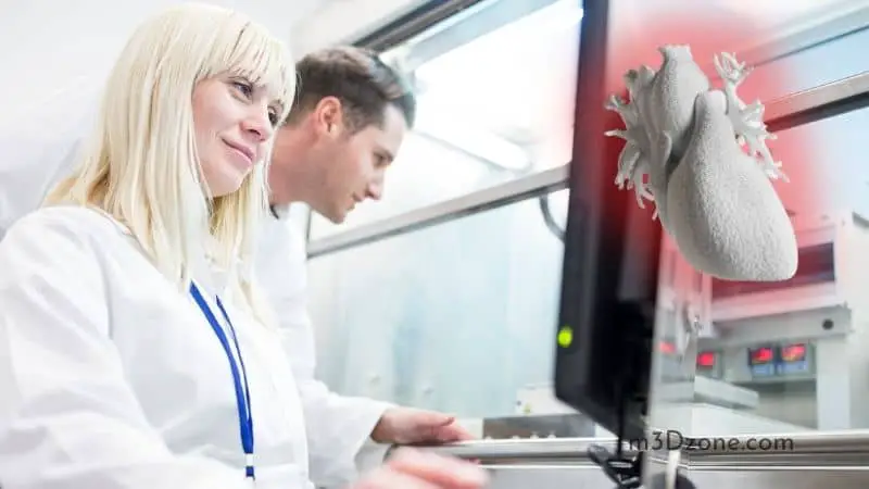
Cardiac Tissue Bioprinting
Cardiovascular disorders have continued to cause mortality in many people. Common heart conditions include cardiac arrest, myocardial infarction, heart failure, angina pectoris, cardiomyopathy, etc. Plus, arteries and veins have shown chronic conditions like stenosis.
The saddening news is cardiomyocytes are irreplaceable as they lack an auto-regeneration or a repair process. Their continual death increases collagen tissue growth, which increases the risk of cardiomyopathy. With these conditions, it’s hard to get a cardiac donor.
But with 3D bioprinting, these health conditions will be manageable. Cardiac tissue bioprinting is challenging because of the complexity of the cardiac muscle, especially in attaining its auto-rhythmic nature.
Cartilage Tissue Bioprinting
Cartilage is a smooth white tissue covering the bone ends. It’s a complex structure made of proteoglycans, collagen, and proteins.
Its outstanding features are that cartilage tissue is avascular, and the nervous and lymphatic systems don’t reach it as well.
Hence, once you continuously injure or cause trauma, you might end up with osteoarthritis or impairment. Tissue engineering currently considers distributing those biological factors through depositing polyethylene and chondrocytes in cartilage tissue bioprinting.
Bone Tissue Bioprinting
Bone tissue is a highly vascularized and structurally complex tissue. Osteo-degenerative and bone fractures may result in injuries and trauma leading to bone tissue dysfunction or a chronic bone defect.
These dysfunctions and defects necessitate bone regeneration which can help restore the damaged bone tissue.
Bone tissue engineering utilizes hydrogels. However, the hydrogels are incapable of forming a mineralized bone matrix.
Thus bone tissue bioprinting promises better results in controllable chemistry and shape maintenance of the tissue integrity.
Which Material Can Be Used in Bioprinting?
The ink must possess your desired biochemical properties, which will help deposit it into the specified patterns. Why?
Because bio-ink facilitates extracellular matrix interactions and cell proliferation and growth. Also, the ink must be biocompatible to support the morphology of the desired tissue.
For cardiac and skin tissues bioprinting, you’ll need a similar bio-ink. You can choose natural polymers like collagen, gelatin, alginate, or hyaluronic acid.
If you prefer synthetic polymers, you could choose poly lactic-co-glycolic acid, polycaprolactone, polyethylene Glycol. Plus, you can choose a blend of synthetic and natural biomaterials.
While selecting bone tissue bio-ink, consider cell specialization, functionality, and cytocompatibility.
You can use gelatin, hydroxyapatite; Gelatin for the preparatory phase and hydroxyapatite to help the printed tissue construct mimic the natural bone tissue.
Wrap Up
3D bioprinting is a tool promising to revolutionize medicine as we know it. From Cardiac tissue engineering to printed bone tissue structures, 3D printing will help face the tremendous challenges in human health.
In conclusion, these tissue engineering applications in cardiac soft tissue, blood vessels, cartilage tissue, etc., will help address the waiting lists for donations and improve health.
We anticipate migrating from conventional cell biology to advanced medicine with three-dimensional tissues and organs very soon.

Recommended Reading
What Is a 3D Printer Operator?
So, what is a 3D printer operator or technician? A 3D printer operator is a competent individual with technical and creative skills in additive manufacturing.
ASA Filament. What Is It Used For?
ASA filament refers to Acrylonitrile Styrene Acrylate. It's a thermoplastic filament used in 3D printing and makes it ideal for engineering and outdoor use.
Why Is It Important to Have a Filament Runout Sensor?
The problem is a lot of printers do not recognize when filament runs out. This is where the filament runout sensor comes into play. Let's find out more!